Electron microscopy is an effective method for assessing the chemical nature of ingredients and provides complementary information for chemical analysis. This type of study applied to food science and technology aims to provide data on the quality of nutrients that are present; detect irregularities in their physical structure; detect the level of processing of components in the food; detect contaminants and microbiological growth; determine fat content; among others.
What is an electron microscope and how does it work?
Electronic microscopes are useful tools in food science. With this type of instrument it is possible to visualize images at higher levels in relation to optical microscopes. In the industry, they are often used to carry out quality control and run process failure analysis. In food technology, it is used for an endless number of applications, including analyzing textures, rheological structures, and biological mechanisms in foods; and contaminants present in products such as fibers, pathogens, etc.
The principle of its operation is based on the use of electrons instead of visible light. Because the wavelength of electrons is much shorter than that of photons, they are used to investigate ultrastructures of a variety of specimens. The electrons are accelerated at high speeds, and impact the sample in a way equivalent to how light could illuminate it, as a result some electrons are reflected by the sample and others pass through it. Images are called micrographs, and they are captured by specialized digital cameras and frame grabbers that detect electrons and build the image.
Types of electron microscopes
There are two types of electron microscopes, the following highlights:
- Transmission electron microscope (TEM): in this type of microscope a fine beam of electrons is accelerated at high speed and conducted to the sample by electromagnetic lenses, so that upon impact of the sample they can pass through and produce a dispersion in different trajectories. The electrons captured by the detector give rise to the high-resolution image, which is magnified and projected onto a fluorescent screen for viewing. This technique is useful to determine details of internal structure in the sample.
- Scanning electron microscope (SEM): the working principle of these microscopes is to sweep the surface of the sample to be analyzed with a beam of electrons accelerated in its electric field by means of a potential difference. Then, using an electromagnet detector, a scan is made through the different points of the surface. In this process of irradiation, part of the initial energy of the electrons is transformed into heat or X-ray emissions, and the emission of secondary electrons also occurs. This type of microscope produces high-resolution, high-depth-of-field images of the sample surface.
Parts of an electron microscope
Electron microscopes consist mainly of:
- Electron source: also called electron gun, it would be equivalent to the light source of an optical microscope. It is a tungsten filament used to generate and direct an electron beam towards the sample. The filament is heated until the energetic level of the electrons in its atoms is such that they can be released and then directed.
- Electromagnetic lenses: In the case of electron microscopes, these lenses produce electric and magnetic fields that allow the path of electrons to diverge or converge. So its function is to deflect the trajectory within the vacuum chamber.
- Vacuum chamber: its function is to prevent electrons from interacting with air molecules, otherwise it is impossible to determine the path of electrons.
- Fluorescent detector or screen: Depends on the type of microscope. If it is a scanning electron microscope, the detector is a sensor whose function is to collect and measure the information of the electrons bouncing off the sample. If it is a transmission electron microscope, the fluorescent screen is responsible for collecting the information of the electrons passing through the sample.
- Sample scanning system: is the integrated system of data acquisition, recording, software and hardware, which is responsible for the operation and control of the microscope, as well as processing the information, analysis and registration of captured images.
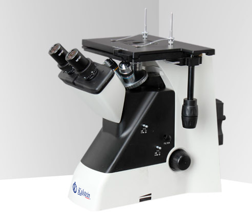
At Kalstein we are MANUFACTURERS, we offer you electronic microscopes with state-of-the-art technology. If you need to know more about our equipment, about PRICE, PURCHASE or SALE, visit us on our catalog HERE