The term Ehlers-Danlos syndrome includes a heterogeneous group of hereditary connective tissue disorders with common characteristics. Patients with this syndrome may present with joint hyperlaxity, soft, hyperextensible skin, abnormal wound healing, and easy bruising. Thirteen clinical subtypes of Ehlers-Danlos syndrome have been detected and the molecular cause of 12 of them is known.
Some symptoms may be common with all types of syndrome and with other connective tissue disorders. Also, because this tissue is distributed throughout the body, each type manifests in varying degrees and in virtually all organ systems. This makes the disorder difficult to diagnose and manage.
An adequate diagnosis of Ehlers-Danlos syndrome is necessary to provide a correct multidisciplinary management of the disease, because currently there are no medical or genetic therapies for this disorder. Management consists of integrated physical medicine and rehabilitation, as well as surveillance for major and specific organ complications. For example, arterial aneurysm and dissection.
Clinical subtypes of Ehlers-Danlos syndrome
This disorder has been classified into 13 subtypes, according to the clinical manifestations they present. Each subtype is mentioned below and is named to describe its most characteristic phenotypic manifestations.
- Classic.
- Vascular.
- Kyphoscoliotic.
- Arthrochalasia.
- Dermatosparaxis.
- Fragile cornea.
- Classic Type.
- Spondylodysplastic.
- Musculocontractural.
- Myopathic.
- Periodontal.
- Cardiac-valvular.
- Hypermobile.
Issues that make the diagnosis of Ehlers-Danlos syndrome difficult
It has been mentioned that this disorder may be underdiagnosed. The diagnosis of Ehlers-Danlos syndrome is considered complex for several reasons, one of them being that many signs and symptoms may be subtle, therefore, medical staff may not be alerted to the possibility of an underlying condition.
On the other hand, the characteristics of Ehlers-Danlos syndrome are similar to the symptoms of other connective tissue disorders. For example, joint hyperlaxity syndrome, Marfan syndrome, or imperfect osteogenesis may occur.
In addition, clinical differentiation between subtypes of Ehlers-Danlos syndrome may be complicated due to overlapping clinical findings. This heterogeneity in the clinical presentation may promote that the diagnosis of this disease is a clinical challenge.
Diagnosis of Ehlers-Danlos syndrome
The diagnosis of this disease has made important advances in recent years. In the early 1980s, the histological characteristics of collagen morphology were used for differentiation of the types of syndrome. Later, the classification of this disorder changed significantly and with it, the diagnostic strategies.
Each subtype of this disease was assigned a set of major and minor clinical criteria, which allows guiding physicians during the evaluation of patients suspected of having Ehlers-Danlos syndrome.
Recently, molecular tests have been used for diagnosis. If a patient meets the clinical criteria for a particular subtype, it should be confirmed with available molecular testing. For the hypermobile subtype, molecular testing is not yet available.
However, molecular analyzes are not always available, so diagnosis is based on clinical findings and use of histopathologic analyzes to confirm the diagnosis and differentiate among some subtypes, for example, vascular, arthrochalasis, and dermatosparaxis.
Use of the tissue processor for the diagnosis of Ehlers-Danlos syndrome
To evaluate histological changes in collagen morphology, adequate tissue preparation is needed, which means chemical preservation or fixation of the material, its support in a solid medium, to be able to cut it into very thin sections and stain it.
Fabric processors allow the material to be fixed, dehydrated and placed in a solid medium. In general, the steps for automated tissue processing are:
- Obtaining the tissue sample: careful handling of the sample is required.
- Fixation: usually, formalin is used. This product needs to penetrate all tissue.
- Dehydration: removal of water from the sample by increasing concentrations of ethanol, in order that it can be infiltrated with paraffin. Failure to do so can lead to problems because paraffin is hydrophobic.
- Clarification: since paraffin and ethanol are immiscible, we must eliminate the latter. Xylene is usually used.
- Paraffin infiltration: done at 60C° with an appropriate histologic paraffin.
- Paraffin block inclusion: the paraffin-infiltrated sample is placed in a paraffin block, allowing the sample to be cut into a microtome.
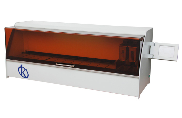
Kalstein tissue processor
At Kalstein we have a wide range of YR series tissue processors for sale. You will find different models that suit different clinical and forensic diagnostic needs. Kalstein equipment, through the all-in-one design, for dehydration and staining of tissue samples, offer reagent savings and guarantee a large space. In addition, they are designed with an LCD screen that facilitates the configuration and display of the parameters of the process being performed. In addition, they have different protocols for dehydration and tissue staining. For more information on Kalstein tissue processors, visit the HERE
We are manufacturers, so in Kalstein you can make the purchase of fabric processors at advantageous prices. For more detailed information, visit HERE


