Rabies is a viral disease that attacks the central nervous system and is characterized by causing acute encephalitis. It is a virus that infects mammals, both domestic and wild, therefore, includes the human being. The virus is found in the secretions or saliva of infected animals, so its mode of transmission is through bites or an open wound on the skin that comes in contact with the saliva of that animal carrier of the disease. Rabies is a dangerous disease that, if not treated promptly, leads to death.
In research on the rabies virus, microscopy has played an essential role in the discovery and description of its structure, as well as its morphogenesis in animals and tissue cultures. The most used microscopes in its characterization and diagnosis are the immunofluorescence microscope and the electron microscope, the importance of these is that it is possible to see very small microscopic agents, impossible to see with the conventional microscope.
Electron microscope in the study of rabies
The structure of viruses can be easily studied by using electron microscopy. With this information, the components and inclusions that make up these agents can be observed in detail. In the case of the rabies virus, this genus belonging to the Rhabdovirus family, when observed under the electron microscope has the shape of a bullet and it is also possible to visualize its nucleus formed by helical RNA single strip.
The electron microscope is a laboratory equipment with which high resolution images can be obtained from biological and non-biological samples. It is widely used in biology and biomedicine, in the detailed study of cells, tissues and viruses, among others, with this can answer questions associated with the structure of these microscopic agents and the basis of many diseases. Its high resolution is due to the use of electrons, which have a short wavelength, rather than the use of visible light, whose wavelength is longer.
Fluorescence microscope in the study of rabies virus
With the DFA or direct fluorescent antibody test, diseases such as rabies can be analyzed, through this test it is possible to verify if the tissues of infected animals have antigens of the virus, which would translate as the presence of the disease. The rabies virus is only present in nerve tissues and not in the blood, as is the case with other viruses, so the tissue to be tested for the presence of antigens is the brain. This test is based on labeling the rabies antibody with fluorescence. The antigen is then incubated with the infected brain tissue to bind to the antibody. Finally, the sample can be observed with an apple-green fluorescence microscope.
Fluorescence microscopes are similar to conventional microscopes that use visible light, except that they have additional fluorescence devices to enhance their capabilities. Conventional microscopes use visible light, which is between 400 and 700 nanometers. On the other hand, fluorescence microscopes should use a higher intensity light source to excite the fluorescent particles in the specimen of interest. Fluorescent particles will emit light of lesser intensity, but with long wavelength, resulting in an enlarged image with bright parts of interest.
Kalstein-branded microscopes
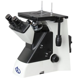
If you are interested in making the PURCHASE of a microscope for your clinical or research laboratory, at Kalstein we are MANUFACTURERS of a wide variety of models at the best PRICES. We have biological, metallurgical, polarization, inverted, stereoscopic, binocular and trinocular microscopes. Our YR series biological models are characterized by: HERE
- Superb picture quality with infinite optical system
- Convenient operation with perfect aerodynamic structure design.
- Better illumination with central sliding capacitor, convenient replacement with phase contrast and dark field capacitor.
To view our microscope catalog you can go to: HERE


