Cervical cancer, cervical cancer or cervical cancer is among the leading causes of cancer mortality affecting women worldwide. It is a type of cancer most often caused by the human papillomavirus (HPV). It is a lesion consisting of malignant cells in the tissues of the cervix, which usually forms slowly over time until the abnormal cells become cancerous, reproduce, and spread to deeper parts of the uterus. Most cases of cervical cancer can be prevented through health programs aimed at detecting precancerous lesions for treatment, including the Pap test and biopsies.
Pre-cancer tests based on the use of a microscope
The Pap test was first suggested by the Greek physician George Papanicolaou in 1928, 15 years later it was accepted by the medical community at large. Then, a 1943 monograph provided a detailed description of how screening should be done. The development of this methodology led him to be the inventor of “Pap smear”, being a pioneer in cytopathology and early detection of cancer.
Screening is done by analysts who examine the sample of cells for signs of malignancy under an optical microscope. This method is used to detect changes in cells caused by the Human Papillomavirus. The sample is taken by a medical specialist using a spatula or brush to gently scrape off cellular material, then placed on a glass slide measuring about 25 × 50 mm. The cells are stained, fixed, and examined. Abnormal samples are processed in a laboratory using a low-power light microscope (colposcope) to observe tissue reflectance. This procedure can not only find evidence of invasive cancer, but also detect cancer precursors, allowing early and effective treatment.
Biopsy is another method of testing often used to diagnose some type of cancer. The aim is to analyze the morphology and structure of cells or tissues in the cervix under a microscope. This test is the only way to determine whether an abnormal area is cancer. A small piece of tissue is removed from the apparently injured area.
Use of the Microscope to Detect Cervical Cancer
High-resolution optical technologies can improve the accuracy and availability of cervical cancer screening, which can identify changes in tissue architecture, cell morphology, and biochemical composition. The microscope provides a diagnosis with high sensitivity and specificity about suspected areas. In addition, histologic techniques such as staining allow the use of dye agents to highlight tissue changes.
Most advanced precancers have vascular changes resulting from the development of new blood vessels. This can be observed and quantified using microscopic image analysis approaches. To visually detect these changes, it is necessary to observe near the limit of optical resolution, since the precancerous lesion can be quite small and local. Therefore, a high-power lens, usually 40x, is used. The projection is initially made at low resolution using a 10x lens, and when something suspicious is seen, the filter changes to 40x.
The types of cervical cancers are classified by how they look when viewed under a microscope. The most common are:
- Squamous cell carcinoma: Squamous cell carcinoma develops from premalignant lesions of the lining of the outer surface of the neck through the formation of many layers of squamous cells.
- Adenocarcinoma: occurs when the epithelium that covers the inner part of the cervical canal, consisting of a single layer of glandular or mucus-producing cells, becomes malignant.
- Malignant tumors: they develop from other cell types resulting in sarcomas, neuroendocrine carcinomas, melanomas, etc.
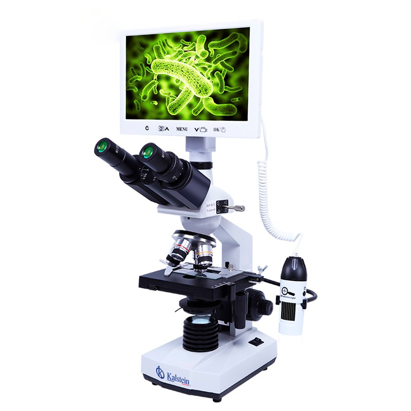
At Kalstein we are MANUFACTURERS, we offer you a variety of types of optical microscopes, with cutting edge technology. If you need to know more about our equipment, about PRICE, BUY or SELL, visit us HERE