A surgical navigation system is a medical team based on the principle of high-precision stereoscopic vision, and which has now become an essential team for the development of computer-assisted surgeries, as they are responsible for guiding the surgeon’s movements during surgery, showing the real-time position of each anatomical instrument and structure. This device allows the use of digital images in surgical procedures, thus providing surgeons with the opportunity to carry out preoperative planning and an effective use of the instruments during surgical procedures.
As for the technologies that use such systems, they can be mechanical, electromagnetic or optical. The most common are optical devices, and these are classified into liabilities or assets. In the former, cameras locate specific markers such as reflective targets, shapes or particular colors, while active systems locate LEDs.
How does surgical navigation technology work?
Surgical navigation technology allows surgeons in the operating room to accurately track the positions of the instrument used and then project the position of the instrument onto preoperative imaging data. This novel technology bears much resemblance to the workings of GPS technology, which allows travelers to see their position on a map.
Before surgery, each patient must have a special high-resolution computed tomography (CT) scan, which is used as a map during the actual surgical procedure. On the day of surgery, that CT scan is loaded onto a computer workstation that processes the images and collects positional data from the tracking system. The first step for any procedure with surgical navigation is a process called “registration,” through which the corresponding points in the patient’s anatomy and preoperative computed tomography are aligned. The register allows calibration of surgical navigation technology.
What is the function of surgical navigation systems?
These surgical navigation systems have as their main function to help accurately locate anatomical structures in open or percutaneous procedures. These systems are used in orthopedics, dentistry, neurology and other surgical specialties. Real-time observations can be made by magnetic resonance imaging, scanner, video camera or other imaging process. Navigation data is incorporated into the image to help the surgeon determine the precise position within the body.
Sometimes medical images are used to plan an operation before surgery. Data integration allows the system to compare the actual position of the target object with the ideal location set during the planning phase. The use of these equipment during different surgical procedures provides greater precision, the performance of less invasive procedures and helps to obtain better surgical results.
What do we offer you in Kalstein?
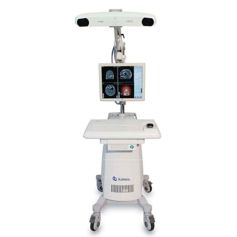
Kalstein is a company MANUFACTURER of medical and laboratory equipment of the highest quality and that have the most advanced technology at the best prices in the market, so we guarantee you a safe and effective purchase, knowing that you have the service of a solid company and committed to health. In this opportunity we present our innovative YR02143 electromagnetic navigation system assisted by computer, which has the following features: HERE
- It is widely used for surgical visualization, planning and navigation to help minimize iatrogenic trauma to surrounding brain tissue and reduce the risk of surgical complications in cranial procedures (such as cranial neurology and ENT surgery).
- The advanced optical tracking system tracks real-time 3D position and orientation of active or passive markers attached to surgical tools, for exceptional accuracy (1.0 mm spatial resolution) and reliability.
- The method of 3D simulation and modeling of anatomical structures in material (such as skin, skull, brain tissue or target injury) can be easily defined by surgical convenience.
- With the navigation probe and advanced optical measurement technology built in, the surgeon can easily quantify the size and position of the lesions, then scientifically design the surgical approach.
- The system provides operators with four navigation modes for comprehensive monitoring of the navigation process.
- The intelligent software will help calibrate and compensate for unexpected changes in anatomical structure and brain change induced by removal of the intracranial lesion area.
- The YR02143 navigation system can operate with a surgeon mouse or a touch monitor mounted on the mobile cart or on the ceiling hanger arm.
- The system automatically stores all patient image data and log information to allow the surgeon to quickly charge and continue surgical navigation against the unexpected power outage.
- It can be used for all neurological and ORL surgeries, especially in deep intracranial lesions, small intracranial volume lesions, small-edge intracranial lesions and minimally invasive surgeries.
For more information we invite you to take a look : HERE


